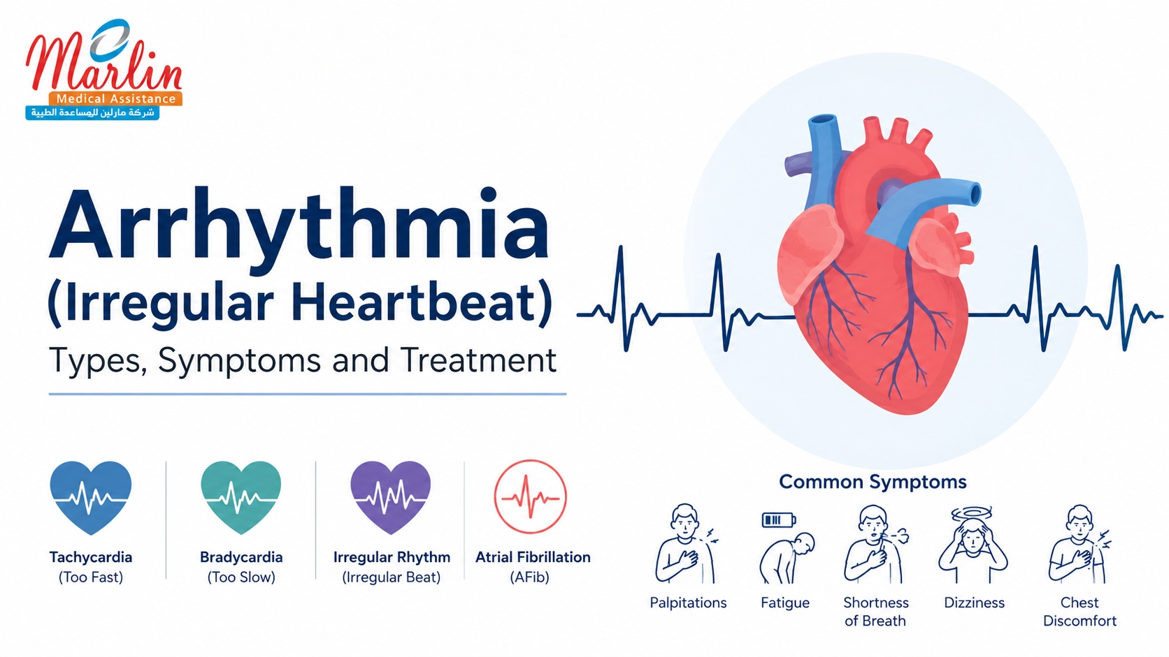
What Is Arrhythmia? — Simple Definition
From a fundamental perspective, cardiac arrhythmia is described as an irregularity from the established normal synchronization of electrical events in the process of heartbeat. While the heart operates as a mechanical pump, it is controlled by a unique intrinsic conduction mechanism. In a healthy body, an electrical impulse referred to as action potential occurs spontaneously in the right atrium due to the sinoatrial node.
The impulse from arrhythmia symptoms travels fast through the working muscle of the atria from the SA node and causes a simultaneous contraction of the atria. This impulse is then met with the atrioventricular (AV) node where there is an important delay of about 120 – 200 milliseconds. This is very important since it helps the ventricles fill completely before starting the contraction process.
After this delay, the impulse moves quickly down through the Bundle of His, divides into two bundle branches, and comes out through the terminal branching network of Purkinje Fibers. This causes a simultaneous contraction of the ventricles from apex to base.
This complex electro-mechanical system gets disturbed due to conditions such as structural heart disease, scarring resulting from ischemia, and metabolic disturbances. The irregularities of the heart rhythms may be generally categorized as follows:
- Tachycardia: A resting heartbeat that is too fast, with more than 100 beats per minute.
- Bradycardia: A irregular heartbeat that is too slow, below 60 beats per minute.
- Irregular Rhythm: The irregularity in the electrical activity of the heart where the heart fails to have a regular beat.
In Swahili-speaking clinical settings within East Africa, especially in Kenya and Tanzania, many patients refer to such disturbing feelings through the use of local descriptive idioms. For example, they could report feeling moyo kupiga haraka ("the heart beating rapidly") or moyo kwenda mbio ("racing heart"). Though these somatic experiences point towards the presence of considerable emotional distress and anxiety, the biological cause of such experiences can be anything from relatively harmless sinus tachycardia to serious re-entrant tachyarrhythmias.
Types of Arrhythmia — AFib, Bradycardia, Tachycardia and More
Heart arrhythmias may be classified by where in the heart the arrhythmia is located (above or below the ventricles), the speed with which the electrical activity is taking place (fast or slow), and the prognosis of the patient.
Supraventricular Arrhythmias
Supraventricular tachyarrhythmias may be initiated by abnormal conduction from the tissue of the atria and/or the atrioventricular (AV) node, which is located above the ventricles:
- Atrial Fibrillation (AFib): This involves electrical abnormality within the atria leading to an irregularly irregular ventricular response.
- Atrial Flutter (AFL): It is related to macro-reentry circuit in the right atrium close to tricuspid annulus and results in fast but regular atrial rhythm (usually 240-400 beats/min) in the form of “sawtooth” waves on ECG.
- Supraventricular Tachycardia (SVT): This term includes paroxysmal arrhythmias such as AV nodal reentry tachycardia (AVNRT), AV reentry tachycardia (AVRT) using
Ventricular Arrhythmias
The Ventricular Arrhythmias can be developed in the contractive ventricular muscle tissue and the subendocardial conducting system:
- Ventricular Tachycardia (VT): Described by the appearance of three or more consecutive premature ventricular complexes at a rapid heart rate (HR>100/min), carrying a high potential for hemodynamic instability.
- Ventricular Fibrillation (VF): The ventricular arrhythmia characterized by the chaotic, very fast activity of the heart muscle resulting in no functioning of the ventricle; without immediate treatment the patient will die within minutes.
- Junctional Ectopic Tachycardia (JET): The arrhythmia that arises from the AV node or His Bundle and most often appears after heart surgery in children.
Bradyarrhythmias
The causes of bradyarrhythmias include the following:
- Sick Sinus Syndrome: Sinoatrial node dysfunction that manifests itself in the form of profound sinus bradycardia, pauses, sinus arrest, and alternating bradycardia and tachycardia.
- Atrioventricular blocks: These arrhythmias vary in terms of the extent of conduction impairment between the atria and ventricles, which includes First-Degree block characterized by the prolonged PR interval of >0.20 seconds, Second-Degree block including progressive prolongation or suddenly stopped beats, and Third-Degree block.
Atrial Fibrillation (AFib) — The Most Common Serious Arrhythmia
Atrial Fibrillation (AFib) is universally accepted as the most prevalent sustained cardiac arrhythmia worldwide. The pathophysiology of AFib symptoms is defined by fast, chaotic and disorganized activation of the atria with atrial rates being anywhere between 350 to 600 beats per minute. Such chaotic electrical activity results in inability for efficient mechanical contraction of the atria.
As a result, the blood accumulates in the left atrium, especially in the left atrial appendage (LAA), resulting in clot formation. In case of release, the clot enters into the circulation system and causes ischemic stroke.
Although the burden of AFib increases worldwide because of the older age population, information obtained from the Kenya Heart Registry proves that patients suffering from this pathology in sub-Saharan Africa are considerably younger than the Western population with median age of 59.0 years. The common comorbidities are hypertension of a longstanding duration with frequent complaints of palpitations, severe fatigue and exercise intolerance.
Dangerous vs. Benign Arrhythmias — How to Tell the Difference
The differentiation between benign and malignant types of arrhythmia is determined by the detailed evaluation of the patient's hemodynamic status and myocardial substrate. Benign arrhythmias such as PACS or PVC are usually found in patients with normally structured heart and do not decrease the cardiac output.
Alternatively, life-threatening arrhythmias impair cardiac output causing significant tissue hypoperfusion. The major clinical manifestations of life-threatening arrhythmia according to the criteria put forward by bodies such as the American Heart Association (AHA) include:
- Syncope/Presyncope: Unexplained episodes of syncope or presyncope due to significantly reduced cerebral blood flow.
- Acute Onset of Severe Dyspnea: Indicating the presence of acute pulmonary edema.
- Isochemic Chest Pain: Present in arrhythmic patients where there is mismatch of oxygen supply to the heart muscles.
- Low Blood Pressure: Less than 90 mmHg indicating hypoperfusion of the body systems.
The clinical significance of arrhythmia is greatly impacted by the presence of structural heart disease. An example is where a single PVC in an individual whose heart lacks ischemic scarring is not life-threatening while in another person whose heart is ischemically scarred due to past myocardial infarction, the PVC can precipitate VT/VF.
Symptoms of Arrhythmia — Palpitations, Dizziness, Fainting
Clinical manifestations of cardiac arrhythmias are extremely diverse, from an entirely asymptomatic condition up to hemodynamic instability. The symptoms that occur in the patient are greatly affected by the heart rate, time of arrhythmia occurrence, patient’s baseline ventricular function, and the perception of the cardiac performance by the patient himself/herself.
Palpitations are the most common symptom of any form of an irregular heartbeat. These symptoms are usually related to rapid heartbeat, heart fluttering, a racing sensation, or the frightening sensation of complete stoppage of the heartbeat.
Within the East African medical setting, these symptoms can be described by employing the indigenous Swahili term such as moyo kupiga haraka or moyo kwenda mbio. One should bear in mind that palpitations can occur in individuals who are having either tachyarrhythmia such as AFib or SVT and bradyarrhythmias.
Dizziness, lightheadedness, and presyncope happen because of the reduction in total cardiac output due to the abnormality in the rhythm, resulting in a temporary decrease in cerebral perfusion. This happens in cases of sustained tachycardias wherein there is limited filling period during diastole, and in the case of severe bradycardias wherein there is a slow rate of flow.
Fainting (syncope) is described as a transient loss of consciousness with recovery. Fainting is considered a high-risk feature in patients with life-threatening arrhythmias. In cases of bradyarrhythmia, it can be a consequence of long-lasting sinus pauses (sick sinus syndrome) or a total heart block (Stokes-Adams attacks). In tachyarrhythmias, syncope happens because of insufficient ventricular filling due to the fast heart rate.
Angina may develop due to fast heart rates especially in people suffering from pre-existing coronary artery disease. The fast heart rate causes reduction of coronary blood flow due to the shortness of the diastole whereas the increased myocardial oxygen consumption leads to myocardial ischemia. Excessive fatigue and decreased physical performance are additional symptoms, which are typical especially in patients suffering from paroxysmal atrial fibrillation.
In addition, many people who have arrhythmia do not show any symptoms at all. This applies especially to paroxysmal and subclinical atrial fibrillation. These "asymptomatic" arrhythmias may be accidentally diagnosed during standard check-ups or, even worse, in case of a patient having a stroke. It highlights the crucial role of screening for such patients having risk factors like long-term hypertension.
Causes and Risk Factors of Arrhythmia
Heartbeat irregularities are hardly ever caused by one specific factor alone. Cardiac arrhythmia usually results from the interaction of several factors including existing anatomical changes in the structure of the heart muscle, metabolic disorders of the body, environmental causes, and even genetic predisposition. Such factors combine to interfere with the normal conduction process of the heart, making the heart to beat either abnormally fast, slow or irregularly.
Structural Heart Disease and Ischemia
CAD and myocardial infarction continue to be the major structural reasons for life-threatening ventricular arrhythmias. The heart attack involves the death of cardiac cells due to the loss of oxygenated blood supply to them.
During the process of healing, the dead tissues get replaced by fibrotic scar tissues. Scar tissue is electrically passive and thus, cannot conduct electricity. Electrical currents are forced to take alternate and long routes through the scar. It makes the electrical milieu ideal for the formation of re-entry loops, and thus, sudden onset of VT or VF.
Other examples of structure changes include cases of dilated or hypertrophic cardiomyopathies, where the structure of the myocardium is distorted, causing the conduction pathway to become elongated. In the same way, heart diseases of the valves, such as mitral stenosis or aortic regurgitation, result in chronic pressure and volume overload, leading to stretching and remodeling of the chamber walls and making the person prone to AFib.
Systemic and Metabolic Factors
Systemic high blood pressure or chronic hypertension is possibly the most common systemic risk factor for arrhythmias in people around the world because it is one of the leading causes of the development of heart disease pathogenesis in sub-Saharan Africa.
Due to chronic increase in systemic vascular resistance, the left ventricle is overloaded and the condition called left ventricular hypertrophy occurs, causing increased pressure in the heart chambers and, consequently, left atrial enlargement and fibrosis. It results in inability of the left atrium to conduct electricity normally and support micro-reentrant waves of AFib.
Changes in endocrine and metabolic status significantly affect the rate and rhythm of the heart:
- Thyroid Diseases: Excessive thyroid hormone production stimulates the receptors responsible for regulating the effect of adrenaline on the myocardium. The result is an increased sensitivity to this hormone and a tendency to develop sinus tachycardia, premature heartbeats, or paroxysms of AF. On the other hand, hypothyroidism slows down the electrophysiological processes and leads to serious sinus bradycardia and conduction defects.
- Electrolytes Balance: Correct content of potassium ions magnesium ions and calcium ions is necessary to preserve the membrane potential of the myocardial cells. Hypokalemia or hypomagnesemia reduces the threshold of depolarization of these cells, which prolongs the duration of action potential and causes after-depolarizations.
Lifestyle Triggers and Autonomic Instability
Short-term exposure to lifestyle stimulants can cause irritation of the electrical system in the heart. Too much caffeine, nicotine, and alcohol are well-known instigators. Alcohol is directly toxic to atrial cells and can cause electrical instability at a local level, which is commonly referred to as "holiday heart syndrome."
Moreover, OSA has been increasingly recognized to be an important inducer of chronic arrhythmias. Episodes of nighttime hypoxia (reduction in the concentration of oxygen in the blood) and significant variations in intrathoracic pressure lead to sudden and enormous activation of the autonomic nervous system resulting in alternating dominance of vagal (which slows the heart rate) and sympathetic (which accelerates the heart rate) influences.
Genetic Predispositions
Genetic channelopathies are relatively uncommon but vital sources of sudden cardiac death in young individuals who have structurally normal hearts. In other words, the problem of genetic mutations consists in structural modifications of the sodium, potassium, or calcium channels within the cardiac muscle and, therefore, affects the action potentials.
Some of the most common examples of genetic disorders include Long QT Syndrome (LQTS), Brugada Syndrome, and Catecholaminergic Polymorphic Ventricular Tachycardia (CPVT). Another important source of postoperative arrhythmias in children is congenital heart disease and surgical scars.
How Is Arrhythmia Diagnosed? — ECG, Holter Monitor, Loop Recorder
A correct diagnosis of an arrhythmia of the heart necessitates proving the arrhythmia by capturing the unusual electrical rhythm. Given that most arrhythmias occur in episodes, physicians employ different methods of diagnosing, which involve different lengths of time for electrical recordings.
The 12-Lead Electrocardiogram (ECG)
The 12-lead electrocardiogram is the definitive first-line test used to diagnose an irregular heartbeat. This involves obtaining an electrocardiogram lasting just ten seconds from twelve angles. Although very useful in diagnosing sustained arrhythmias, such as sustained atrial fibrillation, sustained bradycardia, and acute conduction blocks, the brief nature of the test makes it less useful in diagnosing paroxysmal arrhythmias.
Ambulatory ECG Monitoring (Holter and Event Monitors)
The 12-lead ECG remains the diagnostic gold standard used when there is an abnormal rhythm. It offers a short but comprehensive record of the electrical impulses produced by the heart within a ten-second period. Although it is very effective in making a diagnosis for an arrhythmia that occurs continuously, such as persistent atrial fibrillation, chronic bradycardia or conduction abnormalities, its short time frame limits its usefulness in cases of paroxysmal arrhythmias.
Clinical use of Holter Monitor Kenya in East Africa is common in leading tertiary hospitals. Cardiac services at hospitals like Aga Khan University Hospital Nairobi, The Nairobi Hospital and others make use of this equipment for detecting concealed heart rhythm problems.
Research shows that where a conventional 12 lead ECG is able to detect AFib in only 2%-5% of post stroke cases, the Holter monitor, when used for 24 hours, increases the detection rate. Moreover, cumbersome conventional monitors are gradually being replaced by smaller, wireless adhesive patch monitors that can provide up to 14 days continuous monitoring.
Implantable Loop Recorders (ILR)
When dealing with very rare arrhythmias or recurrent episodes of syncope, whose cause is unclear, the Implantable Loop Recorder (ILR) is considered the best option. The device that looks like a small USB pen is implanted beneath the skin on the chest wall with local anesthesia and records the electrical activity of the heart for up to three years.
Even though the technique is highly efficient, its costs are quite high. The total cost to get an ILR and monitor the patient can be around 3.6 million Kenyan shillings in a year (approximately 300,000 Kenyan shillings in a month).
Diagnostic Availability in East Africa
The state-of-the-art infrastructure for diagnosis has been developing at an incredibly rapid pace. In Tanzania, the most up-to-date cardiac diagnostic services are found at the Jakaya Kikwete Cardiac Institute (JKCI). Being a national training and research institute, JKCI provides comprehensive non-invasive diagnosis that covers 12-lead ECG, Holter monitoring, TEE, cardiac MRI, and a novel 3D electrophysiology mapping system in the catheterization lab.
Treatment: Medications, Ablation, Pacemakers, Defibrillators
Arrhythmias management is very personalized and is dependent upon the specific type of rhythm disorders, clinical symptoms, risk of hemodynamic instability and existence of underlying heart structure abnormalities. In the case of AFib, drug treatment consists of two major lines of treatment approaches: rhythm control and rate control.
- Rate Control: The idea of the rate control therapy is not to restore the rhythm, but only to maintain the heart rate within the physiological limits. The rate is controlled by using beta-blockers (metoprolol, carvedilol) and non-dihydropyridines calcium-channel blockers (diltiazem, verapamil) that suppress the speed of the electrical signal conducted via AV-node.
- Rhythm Control: The rhythm control line includes the use of special anti-arrhythmic drugs (AADs) – Amiodarone, Sotalol, or Flecainide, which affect sodium, potassium and calcium channels to elongate tissue refractory period and decrease conduction velocity.
- Anticoagulation: Since there is a significant danger of a stroke from the turbulent pooling of the blood in the case of AFib, blood clot prevention is critical. The traditional way of doing so involved Vitamin K Antagonists (VKAs) like Warfarin, whose dose needed to be adjusted constantly by testing for INR levels. In contemporary medicine, NOACs/DOACs like Apixaban, Rivaroxaban, and Dabigatran are frequently used because they have a very predictable effect and no need for blood tests.
Pacemaker Surgery — What to Expect
In case a patient suffers from symptomatic bradyarrhythmias such as sick sinus syndrome or complete heart block, a pacemaker must be inserted. Pacemaker surgery has become a well-established procedure, which involves a very low level of invasion and is normally done on an outpatient basis.
The Surgical Process
The procedure is conducted in a specially designed cardiac catheterization laboratory or operating room using a combination of local anesthesia with conscious sedation.
- Incision and Pocket Creation: The surgeon will first sterilize the area and create a 4 to 6 centimeters horizontal incision in the subclavicular region just below the collarbone. Afterward, the surgeon creates a subcutaneous "pocket" by separating the tissues overlying the pectoralis major muscle.
- Lead Insertion and Placement: Using fluoroscopic X-rays in real time, the surgeon inserts one or two insulated electrical lead wires through the subclavian vein into the heart chambers. In a two-chamber device, one lead wire is inserted into the right atrium, while the other is implanted firmly in the right ventricle.
- Testing and Calibration: The leads are attached to an electronic analyzer that determines the electrical stimulation threshold. After testing, the leads are secured on the wall of the heart with either passive tines or an active screw-in system. Finally, the remaining leads are screwed into the pulse generator.
- Closure: The pulse generator is placed inside the pocket created in the chest wall, and then the skin is sutured in layers with absorbable sutures.
Post-Operative and Long-Term Care
Patients have an observation period of a few hours to monitor any problems that may arise, such as a pneumothorax or lead migration. The postoperative care includes safeguarding the wound, keeping the affected arm lower than the shoulder for four to six weeks to avoid tugging at the leads, and undergoing electronic follow-ups. Despite the remarkable longevity of current devices’ batteries, patients will need to have their generators changed locally every seven to twelve years.
Arrhythmia Treatment Available in Kenya
Kenya is one of the major medical centers that treat cardiovascular diseases in East Africa. Diagnosis and management of arrhythmias through electrophysiology studies and implantable devices are done in some of the major tertiary referral hospitals in Nairobi, including the Aga Khan University Hospital (AKUH), The Nairobi Hospital, Kenyatta National Hospital (KNH), and the Moi Teaching and Referral Hospital (MTRH) in Eldoret.
These hospitals have a particular team of experts in interventional cardiology, electrophysiology, and cardiac nursing who can handle structural heart disease. Techniques such as radiofrequency catheter ablation and cryoablation for PVI (Pulmonary Vein Isolation) are among the modern techniques used in these centers.
In addition, full availability of pharmacological therapy is provided across the whole country. Not only traditional drugs like warfarin but also NOACs (novel oral anticoagulants) are provided in order to guarantee stroke prevention. In some cases when patients have complicated congenital or pediatric electrophysiological disorders that demand very specific specialized resources, international healthcare organizations will be ready to provide medical evacuation.
When Is an Arrhythmia an Emergency?
Whereas most palpitations have no risk whatsoever, certain forms of acute arrhythmias can prove a very real and imminent danger in the form of hemodynamic instability, cardiogenic shock, and cardiac arrest, and thus need to be immediately identified in order to ensure the survival of the patient.
Emergency High-Risk Situations
- Sustained Ventricular Tachycardia (VT) and Ventricular Fibrillation (VF): Ventricular fibrillation is described as a medical problem where the electrical coordination of ventricles is completely lost, making the ventricles vibrate and incapable of pumping blood. Cardiac output becomes zero and therefore unconsciousness and cessation of breathing occurs. Sustained ventricular tachycardia may suddenly lead to ventricular fibrillation. Defibrillation using an Automated External Defibrillator (AED) is a must in such cases.
- Third-Degree AV Block / High-Degree AV Block: No electrical conduction from atria to ventricles, resulting in a significantly reduced heart rate of 20-40 beats per minute.
WHEN TO CALL FOR EMERGENCY SERVICES
An arrhythmic heart in combination with fainting is a dangerous cardiovascular emergency. Patients from East Africa may not realize that their palpitations may be an indication of a serious heart disorder. Immediate medical attention is necessary in case an irregular heart rhythm is associated with any of the following conditions:
- Syncope (Sudden loss of consciousness without explanation)
- Crushing pain, pressure, and tightness in the chest
- Terrible dyspnea that develops rapidly
- Extreme dizziness and confusion or cold clammy skin
FREQUENTLY ASKED QUESTIONS
Cardiac Emergency Contact Procedures
In the case of an unstable cardiac emergency, the following procedures should be adhered to by the witnesses:
- In Kenya: Dial the national emergency number 999 for routing of an ambulance.
- In Tanzania: Dial the national emergency number 112, 114, or 115 to quickly transport the patient to a specialist cardiac care institution such as the Jakaya Kikwete Cardiac Institute (JKCI).
- For Emergency Evacuation: If a patient needs to be airlifted from a remote region to a specialized tertiary institution in Nairobi, call AMREF Flying Doctors immediately at +254 20 699 2299.