
What Is Tetralogy of Fallot? — The Four Defects Explained
Learning about your child's having tetralogy of fallot is very scary. This is because it is a particular case of cyanotic heart disease children where there is an actual alteration on how blood flows into the heart and reduces the oxygen level your baby requires. Here, it would be best to understand what is going on within the heart of your precious baby.
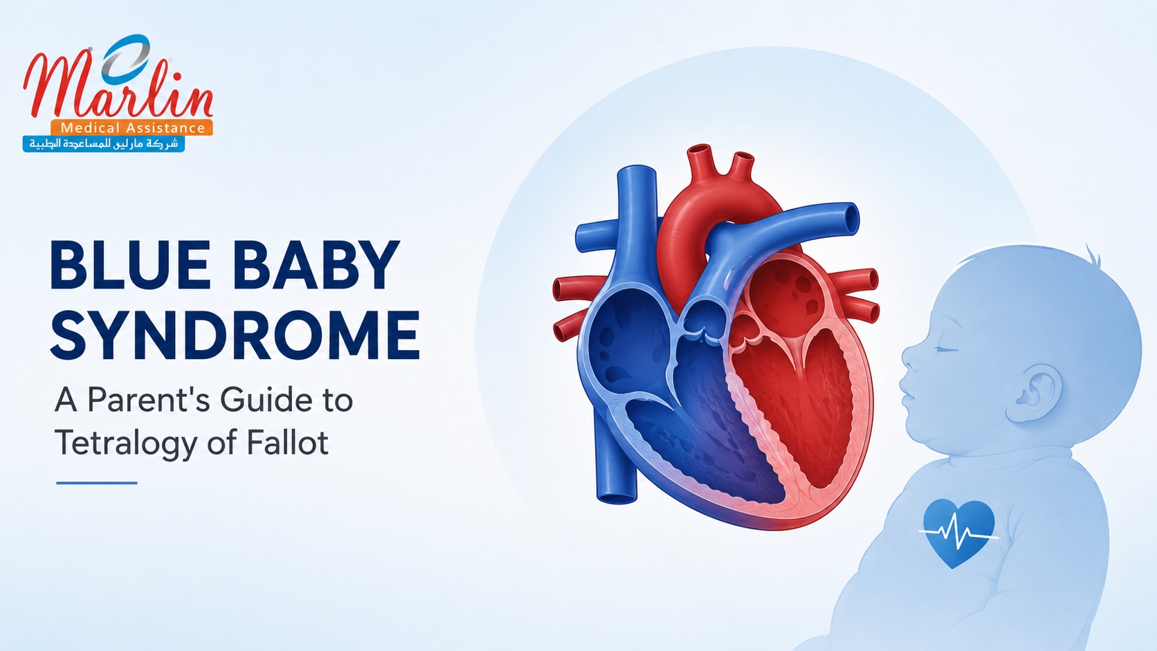
During early embryonic development, a single structural misalignment occurs in the wall that divides the heart's main pumping chambers. This single error creates a chain reaction, resulting in a combination of four distinct, interconnected structural defects. Together, these defects change the heart's plumbing, forcing oxygen-poor blood to mix with oxygen-rich blood.
1. Ventricular Septal Defect (VSD)
In a healthy heart, a solid muscular wall completely separates the left and right lower chambers (ventricles). In babies with tetralogy of fallot, there is a large, non-restrictive hole in this wall. This hole acts like an open bridge, allowing oxygen-depleted blood returning from the body to mix freely with the freshly oxygenated blood returning from the lungs.
2. Pulmonary Stenosis
This defect involves a severe narrowing of the pulmonary valve and the right ventricular outflow tract—the exit pathway leading directly to the lungs. Think of this narrowing as a severe physical bottleneck. Because the pathway is so tight, it restricts the volume of blood that can successfully reach the lungs to receive a fresh supply of oxygen. The tightness of this stenosis is the primary factor that determines how severe your baby's symptoms will be.
3. Overriding Aorta
The aorta is the main pipeline responsible for carrying oxygen-rich blood out to the rest of the body. In a normal heart, it sits squarely on top of the left ventricle to distribute pure, oxygenated blood. In this condition, the aorta is shifted to the right and hovers directly over the VSD hole. Because of this faulty positioning, the aorta straddles both chambers, collecting and distributing a mixed, poorly oxygenated stream of blood throughout your child's body.
4. Right Ventricular Hypertrophy
Due to the narrowness of the pathway to the lungs, there will be increased pressure on the right ventricle to contract forcefully enough to push the blood past the blockage. The prolonged pressure leads to thickening of the muscle wall of the right ventricle. This thickening of the muscle is a result of adaptation by the body but limits its efficiency in the end.
When these four defects interact, they severely disrupt the body's normal oxygen balance. Instead of sending all oxygen-poor blood to the lungs and all oxygen-rich blood to the body, the heart is forced to circulate a mixed, low-oxygen stream. This systemic oxygen deficit is what ultimately leads to the visible physical signs of blue baby syndrome.
Why Do Babies with ToF Turn Blue? — Cyanosis Explained
That is why blue baby syndrome has a physical appearance of the baby which is blue or purple and that makes it frightening for parents to see. It is known medically as the central cyanosis and although frightening, having an idea on what causes it will make it easier for you to cope.
There are two separate systems in the heart. One is where blood from the right ventricle is pushed to the lungs and in a relatively lower pressure while another where blood is sent to the rest of the body in very high pressure. In case of an infant having tetralogy of fallot, the pulmonary valve is highly narrowed.
This resistance forces the right ventricle to push with extreme effort, causing the blood pressure on the right side of the heart to skyrocket until it matches or exceeds the pressure on the left. This pressure inversion forces oxygen-poor blood—which has just returned from the body and desperately needs to visit the lungs—to take a dangerous shortcut. It flows directly across the ventricular septal defect (VSD) into the left ventricle and straight up into the overriding aorta.
This particular type of blood bypasses the lungs; therefore, it does not receive any fresh supply of oxygen. The deoxygenated blood is naturally dark and bluish in color as opposed to the bright crimson red blood that has oxygen. The presence of this poor oxygen supply when circulating within the baby’s body results in reduced oxygen content in the body as a whole. The oxygen deficiency becomes noticeable in regions where the skin is thin and there are many blood vessels.
This baseline cyanosis can suddenly worsen during what is known as a hypercyanotic episode, or a "Tet spell". These spells are sudden, acute crises of profound oxygen depletion. They are typically triggered by everyday activities that cause physical strain or stress, such as crying, feeding, passing stool, or even waking up quickly in the morning.
When a child suffers from Tet spell, there occurs an involuntary contraction of muscle fibers that lie just beneath the pulmonary valve, which almost shuts down the passage to the lungs. Almost all of the blood deprived of oxygen gets shunted to the rest of the body bypassing the lungs, turning the baby’s face into blue and forcing him to breathe heavily.
Signs and Symptoms of Tetralogy of Fallot in Newborns and Infants
As a parent, learning to recognize the signs of congenital heart disease cyanosis is essential for keeping your baby safe. While some infants with mild narrowing of the pulmonary pathway may look relatively pink at birth, most babies with tetralogy of fallot will begin to show noticeable physical symptoms within their first few weeks of life as their body's metabolic demands increase.
The most common symptom is cyanosis, which refers to the blue appearance caused by low oxygen in the blood. At first, this discoloration will be intermittent. You will find that your baby shows bluish tinge of skin near the mouth, eyes, or fingernails only when engaged in activity such as crying, eating, or passing stools. As the condition progresses and the strain on the heart becomes continuous, the bluish tint could advance to central cyanosis, where the lips, tongue, and other mucosal areas appear dusky blue continuously.
The act of feeding needs an enormous amount of physical energy for the baby. The reason why the heart which is already not healthy enough will be unable to satisfy such great metabolism is that the babies get very tired from feeding through breast milk or from bottles. Your baby may start drinking some milk, sweat excessively, breathe quickly, and after a while stop because he/she simply cannot go on anymore. They are unable to complete their feedings; thus, they are often diagnosed with insufficient weight gain and failure to thrive.
Clubbing – swelling and rounding of fingers and toes due to chronic hypoxia of tissues – is one of the common symptoms which develop in babies who have lived for more than 6 months without undergoing surgery.
Additionally, older toddlers who are walking will instinctively adapt to their condition through a unique behavioral trait: during physical play or when they begin to feel short of breath, they will suddenly drop into a deep squatting position.
The Squatting Mechanism: As one bends their knees and hips, the large arteries in the lower body become compressed. Compressed structure causes resistance in the arterial system of the body, which creates artificial increase in the blood pressure in the left side of the heart. It is this change in the blood pressure that counters the left-to-right shunt and redirects more blood to the lungs.
TET SPELL EMERGENCY CAREGIVER PROTOCOL
"Blue Spell" or "Tet Spell": A hypercyanotic "blue spell" is an acute emergency situation which demands prompt physical intervention. If your child's skin suddenly turns extremely blue, if he/she struggles to breathe, gets unusually irritated, or loses his/her consciousness, here's what you need to do right away:
1. Apply the Knee-to-Chest Mechanism: Take your baby in your arms and fold his/her knees against his/her chest in order to hold him/her in such a position. In case of an older child, make sure he/she squats deeply and tightly. Structural changes cause increased systemic vascular resistance and change the pressure balance in the heart, thus redirecting blood supply to the lungs.
2. Calm Your Child Down: Fear and panic cause intense spasms in the muscles situated under the pulmonary valve and prevent oxygen supply to the lungs. Hold your child close, calm down and comfort him/her.
3. Rush to the Hospital: In the meantime, take your child to the hospital for further examination and treatment.
Diagnosis — When and How ToF Is Detected
It becomes a life-changing experience when you find out that your child is suffering from tetralogy of fallot, and sometimes the time of this experience largely depends on the availability of medical screening options that one may have. In countries where there are sophisticated health care facilities, about half of the children diagnosed with this condition are identified before they are even born. The reason is that this is done through fetal echocardiography which means the ultrasound of the baby's heart that can be done during pregnancy at 18-22 weeks.
This early knowledge provides the family with some valuable advantage. They have the chance to prepare themselves mentally for the diagnosis and seek advice from pediatric cardiologists.
Routine Newborn Screenings
Babies with congenital heart disease that has been undiagnosed during pregnancy are discovered through regular neonatal examinations. The pediatrician listens to their heart within the first 24 to 48 hours from birth using a stethoscope. In this particular case, there is always a very audible and harsh systolic ejection murmur in the area of the left border of the breast bone of such a baby. It is caused by very strong turbulence resulting from the passage of blood through an obstructed pulmonary path.
Along with a murmur detection, modern hospitals also apply newborn pulse oximetry screening, which is a very quick procedure. It consists of the application of a little sensor onto the baby’s foot or hand that will detect the initial level of oxygen in the blood. This test helps to detect an invisible deficit of oxygen in the blood of a newborn baby who can be very pink when sleeping in the nursery.
The Definitive Diagnostic Tool
If a murmur or low oxygen reading is detected, a pediatric cardiologist will immediately order a transthoracic echocardiogram. This is the definitive, gold-standard tool used to confirm the diagnosis. The painless test uses advanced ultrasound waves to map out the heart’s internal architecture in real-time, allowing the specialist to visually measure:
- The exact size and placement of the ventricular septal defect (VSD).
- The degree of tightening in the pulmonary valve.
- The precise positioning of the overriding aorta.
In low-resource environments like rural Kenya or rural Tanzania, prenatal ultrasounds and screening tests after birth may not be available. In such a population, diagnosis takes place several months or even years later. The disease usually goes unnoticed until the child exhibits severe physical symptoms at home like blue skin tone during feeding, constant fatigue, poor weight gain, or a frightening "tet spell" during crying spells. Although the late diagnosis indicates that there has been considerable stress on the heart, an accurate echocardiogram continues to be the first essential step towards fixing the problem.
Treatment — Open Heart Surgery for Tetralogy of Fallot
While a diagnosis of cyanotic heart disease children face sounds frightening, it comes with an extraordinary message of hope. The definitive, curative blue baby treatment is open-heart surgery. Thanks to decades of advancements in pediatric cardiothoracic surgery, corrective procedures completely restructure the heart, allowing the vast majority of children to live full, active, and long lives.
For most infants, a complete intracardiac repair is performed between 3 and 6 months of age. Performing the surgery during this window prevents the heart muscle from sustaining long-term damage from chronic oxygen deprivation and high pressure.
What a Complete Intracardiac Repair Involves
The total repair procedure is an extremely complicated surgery that is carried out using general anesthesia. The procedure involves a median sternotomy cut along the breastbone and the utilization of cardiopulmonary bypass equipment. The equipment performs the functions of the heart during the procedure by temporarily taking over the task of circulating oxygenated blood while the heart is stopped and at rest. In the process of the procedure, there are two important repairs done:
- Blocking the VSD: The surgeon will place a synthetic patch (usually composed of Dacron or Gore-Tex) over the hole in the heart’s ventricle. This will block all holes completely and prevent the mixing of oxygen-rich and oxygen-poor blood.
- Expanding the Pulmonary Passage: The surgeon will cut off any muscle bundles that are causing a blockage and widen the narrow valve and passage by use of a transannular patch. This will remove the bottleneck, enabling blood to pass from the right ventricle to the lungs without any hindrances.
Now that the hole is blocked and the passage is expanded, the overriding aorta will pull blood only from the left ventricle, resulting in a normal blood circulation process.
The Staged Surgical Approach (Palliative Shunt)
This is not possible in all newborns. Premature delivery, extreme smallness, and the presence of anatomically challenging factors such as a totally occluded pulmonary valve make staged surgery a necessity.
In such fragile patients, doctors would perform a life-sustaining palliative operation within days after delivery known as a modified Blalock-Taussig-Thomas (mBT) shunt. In this process, surgeons would place a small artificial tube between a vessel in the subclavian artery and the pulmonary artery. The mBT shunt serves as a temporary bypass which ensures consistent blood flow to the lungs, allowing the patient to stay alive until the intracardiac surgery.
Post-Operative Recovery and Outcomes
The post-surgical recovery process of CHD surgery infant suffering from CHD occurs within a Pediatric Intensive Care Unit (PICU). The newborns are put on a ventilator machine for breathing support for 24 to 72 hours. Medical personnel keep an eye on cardiac rhythm and blood pressures of babies using small lines.
Once the child is breathing on their own and their vital signs stabilize, they transition to a general pediatric ward. Most families are able to take their baby home within 7 to 14 days after surgery. The immediate physical change is nothing short of miraculous—your child’s skin, lips, and fingers will turn a healthy, vibrant pink as their body finally receives the rich oxygen supply it deserves. In well-resourced medical centers, the survival rate for this complete repair now exceeds 95%, offering an incredibly bright future.
ToF Surgery in East Africa — What Is Available?
Being diagnosed with tetralogy of fallot is very stressful for anyone; however, knowing how to access reliable healthcare services is even harder for families residing in various parts of East Africa. The medical community may have achieved great strides through innovation and discoveries; nevertheless, the region is very poorly equipped to handle complicated open heart operations for many children who have congenital heart disease.
The Reality of Regional Surgical Capacity
Access to modern pediatric cardiac facilities is a major problem in Sub-Saharan Africa. According to figures collected by the World Health Organization (WHO), although only 1% of all newborn babies in the world suffer from CHD, fewer than 10% of those suffering from the condition in Sub-Saharan Africa can receive lifesaving surgery.
If we consider the specific type of cyanotic heart disease children are affected by, the figure for the number of tetralogies of fallot is around 8% to 11% of the total number of regional CHD. In comparison to the high regional number of births, the disparity is brought into focus:
| Kenya | ~1,293 cases per year | ~120 to 150 operations annually |
| Tanzania | ~2,620 cases per year | ~250 operations annually |
| East Africa (Combined) | ~4,950 cases per year | ~370 to 400 operations annually |
Sourced Data (WHO / JKCI Registry 2025): The vast majority of the nearly 5,000 children born with ToF each year in East Africa are left on waiting lists because local infrastructure can only accommodate a tiny fraction of all congenital cases.
Infrastructure Challenges in Kenya and Tanzania
The infrastructure challenges facing publicly funded tertiary hospitals such as KNH in Kenya are many. For instance, there are no special pediatric cardiac intensive care beds for children who have had ToF. As a result, surgeries have to wait until there is an availability of an ICU bed. Although there are very skilled surgeons in private hospitals such as Nairobi Hospital and Mater Hospital to carry out ToF surgery in Africa, it is too costly.
In Tanzania, the Jakaya Kikwete Cardiac Institute (JKCI) in Dar es Salaam has made incredible strides since its founding, expanding its capacity to perform around 250 pediatric surgeries a year. However, because the demand heavily outpaces the slots, children are often operated on much later than the ideal 3-to-6-month window. A 2025 facility-based study at JKCI revealed that the median age for ToF surgery was 3 years old, and over 61% of these pediatric surgical candidates presented as severely underweight due to the prolonged metabolic strain of chronic oxygen deprivation.
The Lifeline of Medical Evacuation
Since any delay in blue baby treatment would increase the probability of developing severe tissue hypoxia as well as dangerous Tet spells, evacuation of East African families to specialized international centers is now an essential requirement. India stands out as the preferred location, due to its high-quality pediatric cardiothoracic surgeons, specialized ICUs, survival rate exceeding 95%, and cost effectiveness compared to Western hospitals.
This is where dedicated medical facilitation networks like Marlin Medical Assistance step in to bridge the gap. They manage the entire medical journey—from initial clinical consultations and fast-tracking medical visas to arranging flights and securing affordable care packages at premier Indian pediatric cardiac centers—ensuring your child bypasses local waitlists and receives the immediate, expert care they deserve.
Life After ToF Surgery — What Parents Should Know
With the completion of your child's tetralogy of fallot Kenya or East Africa treatment process, the future looks bright for your little one. There is a great feeling of peace and relaxation knowing that your baby's skin color changes from pale blue to a healthy pink color. In fact, the majority of children have a quick “catch up” growth spurt, where there is an instant burst of energy and hunger.
Your little one will be able to attend school and play sports like any other child. However, it is essential to know that surgery is a correction and not a cure. This means that your child will have regular visits to a congenital cardiologist for heart examinations. The most common problem for adult tetralogy of Fallot patients is the leaking pulmonary valve.
Because of this, many patients need a pulmonary valve replacement procedure when they become adolescents or adults. With today's advances in medicine, many of these additional valve procedures can be done in non-invasive transcatheter techniques without the necessity of undergoing open-heart surgery again.
Get your consultation from Marlin today
Medical Disclaimer: This guide is for informational purposes only and does not substitute for professional medical advice, diagnosis, or treatment. Always consult a qualified pediatric cardiologist or healthcare provider regarding any questions about a medical condition.